If non-accidental head injury (or shaken baby syndrome) is suspected, it is likely that an ophthalmologist (specialist eye doctor) will examine your child’s eyes to see if there are any retinal haemorrhages present. Retinal haemorrhaging is one of the three signs of the ‘triad’.
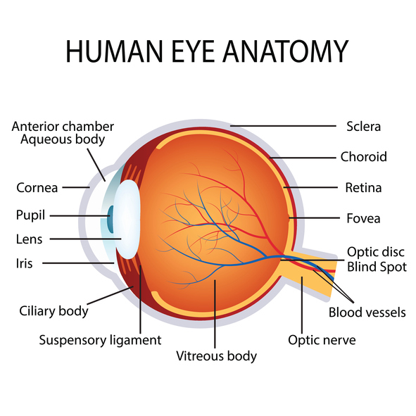
The picture above shows the eye from the side and labels the parts that you are most likely to come across within an expert report.
The retina is a structure of multi-layered tissue at the back of the eye. Retinal haemorrhaging is basically bleeding into the retina within the eye. Features that make retinal haemorrhaging more severe can include whether they are present in one or both eyes, the number of haemorrhages, whether they are in every layer of the retina or whether they extend to the outside of the retina or are just focused around the centre.
Retinal haemorrhaging can be caused in many ways including direct trauma to the eye, raised intravascular pressure, raised intracranial pressure, raised central venous pressure or interaction with the vitreous jelly caused by the effects of shaking.
Research has found that retinal haemorrhages are more commonly caused in non-accidental injury than in an accident. It is well established that retinal haemorrhaging can occur at birth but in most cases these haemorrhages will resolve within 4-6 weeks.
