The brain is a complex organ. The most common intracranial injury that can arise in allegations of non-accidental injury is a subdural haemorrhage. It may help to understand a little about how the brain is formed…
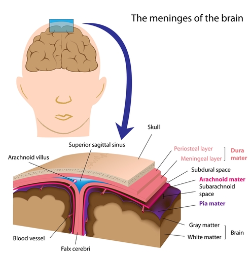
The brain is enveloped in three layers – the pia, the arachnoid and the dura. Between the pia and the arachnoid there is a space (the sub-arachnoid space) which is filled with cerebrospinal fluid. Between the arachnoid and the dura there is no actual space (it is often referred to as the subdural space which is misleading); rather, there is the potential for a space to be created if fluid or blood leaks into it.
Subdural haemorrhage refers to blood in the potential space between the arachnoid and the dura. Subdural haemorrhages are associated with trauma. They do not occur spontaneously in children and so, in the absence of a bleeding disorder, some pre-disposing malformations or rare metabolic disorders, medical opinion is that they are caused by trauma (the allegation usually being shaking with or without an impact). It is thought that the veins connecting the brain to the skull (bridging veins) are torn and this is what causes subdural haemorrhages or subarachnoid haemorrhages.
Bleeding on the brain cannot be dated with precision. Fresh blood is referred to as an acute bleed and refers to blood that is less than 10 days old radiologically. Sometimes the clinical presentation (behaviour) of a child is of more assistance when trying to pin down a timeframe for subdural haemorrhage.
Occasionally, fresh blood fails to absorb back into the brain and become old blood, perhaps with membranes within the blood. This is known as a chronic subdural haemorrhage (old; over two weeks). An infant’s skull bones are supple and not yet fused; this means that the skull often expands where there is a chronic subdural haemorrhage in an attempt to relieve the pressure on the brain. As a result, the child may not have any symptoms of brain injury other than a slightly larger head (often unnoticed by carers). It is possible for there to be re-bleeds into a chronic subdural haemorrhage.